| TITLE |
|---|
| AUTHORS |
| SOURCE |
(DOI: 10.1109/TNS.2006.882795)
|
| ABSTRACT |
|
An 8-layer depth of interaction (DOI) detector was designed based on the technique we have developed for 4-layer DOI detectors. The new detector achieves 8-layer DOI encoding by pulse shape discrimination (2-layer DOI encoding) and an optimized reflector arrangement in a 3-dimensional crystal array (4-layer DOI encoding). Its capability was proved with an 8-layer 10 x 10 Gd2SiO5 (GSO) crystal array coupled to a 256-channel flat panel position sensitive photomultiplier tube (256ch FP-PMT). The dimensions of each crystal element were 2.90 mm x 2.90 mm x 3.75 mm and the interval between 16 x 16 multi anodes in the 256ch FP-PMT was 3.04 mm. Two different dopant concentrations in GSO were chosen for pulse shape discrimination: GSO crystals of 0.5 mol% and 1.5 mol% Ce dopant that have 60 ns and 35 ns scintillation decay times, respectively. In the crystal array, Layer-1, the farthest from the 256ch FP-PMT, and Layers-3, -5 and -7 were composed of GSO crystals of 0.5 mol% Ce dopant. All other layers were composed of GSO crystals of 1.5 mol% Ce dopant. Performance of the 8-layer DOI detector was evaluated by irradiating with 662 keV uniform gamma-rays and the capability was judged to be good. To prove the validity of the layer encoding, a fan-beam of 662 keV gamma-rays was irradiated onto the side face of each layer. The obtained 2-dimensional position histograms showed the right structure in each corresponding layer clearly. Light output uniformity among DOI layers and energy resolutions were measured on the pulse height distributions of the central crystal in each layer. The crystals in Layer-2 and Layer-3 showed the smallest light output and their full energy peak channels were 62% to the largest peak channel of the Layer-8 crystal distribution. Energy resolutions were about 15% for all eight layers.
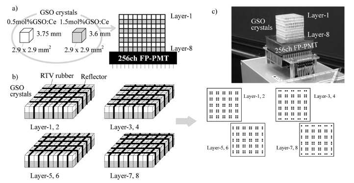 Fig. 8. Fig. 2. (a) Structure of an 8-layer DOI detector for preliminary experiments. It was composed of a 10 x 10 x 8 array of GSO crystals of two different dopant concentrations and a 256ch FP-PMT. (b) Reflector arrangement in the 8-layer DOI detector and diagrams of 2D position histograms produced with the reflector arrangement. c) 8-layer DOI detector used in the measurement. |
| KEYWORDS |
| depth of interaction (DOI), GSO, positron emission tomography (PET), PET detector module, resolution, block, design |