| Tomoaki Tsuda, Hideo Murayama, Keishi Kitamura, Naoko Inadama, Taiga Yamaya, Fumihiko Nichikido, Manabu Hamamoto, Hideyuki Kawai, and Yusuke Ono |
|
Previously, we proposed a new depth of interaction (DOI) encoding method and proved that it worked successfully with four-layered Gd2SiO5 crystals for a small animal positron emission tomography (PET) detector. We are now planning to develop a small animal PET scanner, jPET-RD (for rodents with DOI detectors), which has both high resolution and high sensitivity by the use of a DOI detector with a 32 x 32 x 4 crystal array. The scintillator for the detector will be Lu2(1-x)Y2xSiO5 (LYSO). In this work, we evaluated performance of a DOI detector composed of four layers of a 12 x 12 LYSO (Lu: 98%, Y: 2%) crystal array by irradiating 511 keV gamma rays uniformly. The new encoding method was used for crystal identification. The size of each crystal was 1.46 mm x 1.46 mm x 4.5 mm. The crystal block was coupled to a 256-channel flat panel position sensitive photomultiplier tube, which has 16 x 16 multi anodes at intervals of 3.04 mm. As we expected, all crystals are expressed on a single two-dimensional position histogram without overlapping. Energy resolution of all events is 21.8% and time resolution of all events is 0.69 ns in FWHM. When layers are counted from the top, the energy resolutions of the first, second, third, and fourth layer events are 11.6%, 12.3%, 13.3%, and 19.1% and the time resolutions are 0.60 ns, 0.59 ns, 0.60 ns, and 0.66 ns, respectively.
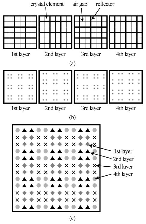
Fig. 3. (a) Reflector arrangement (top view). (b) Pattern diagrams of the 2-D position histogram, corresponding to the crystal array with the reflector arrangement (a). (c) Pattern diagram of the 2-D position histogram which will be obtained with the for-layer DOI detector. Each circle, triangle, cross and diamond corresponds to the peak of a crystal element in the first, second, third and fourth layers, respectively.
|