| TITLE |
|---|
| AUTHORS |
| SOURCE |
(doi:10.1063/1.277715)
|
| ABSTRACT |
|
We introduce experimental systems which use accelerators to evaluate scintillation properties such as scintillation intensity, wavelength, and lifetime. A single crystal of good optical quality is often unavailable during early stages in the research and development (R&D) of new scintillator materials. Because of their beams’ high excitation power and/or low penetration depth, accelerators facilitate estimation of the properties of early samples which may only be available as powders, thin films, and very small crystals. We constructed a scintillation spectrum measurement system that uses a Van de Graaff accelerator and an optical multichannel analyzer to estimate the relative scintillation intensity. In addition, we constructed a scintillation time profile measurement system that uses an electron linear accelerator and a femtosecond streak camera or a microchannel plate photomultiplier tube followed by a digital oscilloscope to determine the scintillation lifetimes. The time resolution is approximately 10 ps. The scintillation spectra or time profiles can be obtained in a significantly shorter acquisition time in comparison with that required by conventional measuring systems. The advantages of the systems described in this study can significantly promote the R&D of novel scintillator materials.
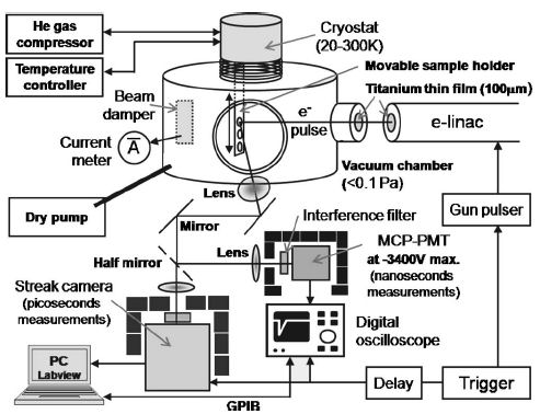 FIG. 2. Schematic diagram of the scintillation time profile measurement system using an e-linac and a femtosecond streak camera or a MCP-PMT followed by a DOS. The streak camera is mainly used for measurements of lifetimes shorter than 1 ns, and the MCP-PMT is used for the measurements of lifetimes longer than that. The accelerator and detectors share the trigger signal. They are placed in a dark room and are controlled from the operation room. |